Introduction
Post-operative infections in patients undergoing living donor liver transplantation (LDLT) are a major cause of morbidity and mortality in our center and elsewhere [1]. Patients are at the highest risk of acquiring infections during the first post-operative month. These include both surgical site infections and remote infections involving the chest, urinary tract and bloodstream [2]. Such infections are usually masked due to immunosuppressive therapy which dampens the clinical and laboratory parameters of the inflammatory reaction. This leads to a delay in diagnosis and hence an undesirable outcome [3]. A myriad of risk factors are known to increase the likelihood of infections in transplant patients, but the risk of infection for individual patients remains unpredicted.
The study aimed to develop a practical and efficient prognostic index for early identification and possible prediction of post-transplant infections using risk factors identified by multivariate analysis. Discrimination of patients according to infection risk is vital in determining the tricky balance between over- and under-immunosuppression. Also, high risk patients should undergo more aggressive prophylactic and empirical chemotherapeutic measures.
Material and methods
Study design and study sample
The cohort study included 100 Egyptian patients with end stage liver disease (ESLD) who underwent LDLT during the first phase of introducing this procedure in our center. This study was conducted at Kasr Al Ainy Hospital, Liver Transplantation Department, Faculty of Medicine, Cairo University. Adult patients were consecutively enrolled in the study from 2004 until 2009. All of them had liver cirrhosis due to chronic HCV genotype 4 infection. Indeed, this is the most common cause in this age group, ranging from 20 to 63 years. The study examined the early post-operative period for LDLT recipients, during the first post-operative month.
The study included 90 males and 10 females. All patients who fulfilled the following inclusion criteria were enrolled: post-hepatitic cirrhosis, HCV positive, genotype 4, Child B/C with a MELD score of 13–25.
Patient management and decisions were independent from the study and totally subject to our center’s protocol. Accordingly, thorough clinical examination, and laboratory tests were done twice daily during the first 5–7 days in the intensive care unit and then once daily during the rest of the hospital stay on the ward. Surviving patients are under regular follow-up.
Data collection and management
More than 160 fields per patient were documented including pre-operative, operative and post-operative data. The medical team was blinded to all research data. Data included patient history, clinical, laboratory, and ultrasonographic information as well as immunosuppressive and antibiotic regimens, complications and outcome. Data were entered using Microsoft Excel at the transplantation unit.
Standard immunosuppression and antibiotic prophylaxis
All patients received intravenous piperacillin/tazobactam, oral nystatin and oral acyclovir during the first post-operative week.
First line immunosuppression included corticosteroids, tacrolimus and mycophenolate mofetil. Acute rejection episodes were treated with corticosteroid pulse; one patient received antithymocyte globulin (ATG). Second line drugs included cyclosporine, sirolimus, and everolimus.
Outcome measures
Diagnosis of infection
Clinical suspicion of infection was considered when one or more of the following were present: fever, leucocytosis, neutrophilia showing shift to the left, rise in CRP, relevant X-ray findings and positive culture from any body fluid. Fever was defined as any elevation in body temperature beyond normal diurnal variation.
The definition of bacterial infection was based on the criteria of the Centers for Disease Control and Prevention (CDC) [4, 5] as specific changes combined with clinical symptoms such as fever, cough and hypoxemia confirmed by at least one positive blood culture or imaging results (X-ray, MRI, CT).
We attempted to identify all infections which occurred after transplantation and categorize them according to time after transplantation, site of infection, type of pathogen, severity, and outcome. Strict criteria were employed to define infections included in this study. They are as follows:
Bacteremia
Isolation of Listeria monocytogenes, Staphylococcus aureus, Candida and aerobic Gram-negative bacilli in one blood culture. Other bacteria require, in addition, a confirmatory blood culture or a positive culture from an infection site.
Elevated C-reactive protein (CRP) was in favor of bacterial infections [6].
Soft-tissue infection
Isolation of causative agent from a soft tissue site showing signs of infection requiring surgical management.
Peritonitis
Neutrophil count in diagnostic peritoneal tap ≥ 300, with bacterial isolation by fluid culture. The presence of an infective intra-abdominal collection is diagnosed by ultrasound or found at laparotomy.
Cholangitis required
Fever, cholestatic pattern in liver function tests and a positive culture from bile duct secretions.
Pneumonia
New relevant findings in chest X-ray or CT chest, typical symptoms such as productive cough, dyspnea or evidence of hypoxemia. Direct microscopic examination of bronchoalveolar lavage is used to identify bacterial (especially Pneumocystis spp.), and fungal pathogens using appropriate stains. Isolation of causative agent by culture of sputum or bronchoalveolar lavage is otherwise required.
Urinary tract infection
Lower urinary tract infection is diagnosed by the presence of pyuria with positive urine culture, with or without dysuria and frequency. Upper urinary tract infection is diagnosed, in addition to the former criteria, by fever, flank pain or positive blood culture isolation of the same pathogen.
Cytomegalovirus (CMV) disease
Fever ≥ 38°C for 1 week with atypical lymphocytosis, leucopenia or thrombocytopenia. Confirmation is made by CMV PCR. Tissue involvement was diagnosed by histologic presence of inclusion bodies or direct culture.
Disseminated fungal infection
Clinical picture consistent with sepsis together with isolation from blood cultures or antigens in blood or biological fluids for candidiasis, aspergillosis or cryptococcosis.
Procalcitonin was used in selected cases where the diagnosis of infection could not be determined in spite of the strong clinical suspicion as a marker of bacterial infection. It has also been shown to be of prognostic value after liver transplantation [8]. However, it is not practical to be applied, especially in this setting where daily evaluation is required in the early post-transplant period, due to its high cost and limited availability in our center.
Informed consent and ethical considerations
Written informed consent was obtained from all subjects. The study protocol was revised and accepted by the faculty ethical committee.
Predictors of infection
Potential predictors of infection were chosen based on a review of the literature. Pre-operative predictors included age, sex, weight, height, body mass index (BMI), blood group, diabetes, hyperlipidemia, hypertension, MELD score, Child score, spontaneous bacterial peritonitis (SBP), hepatic coma, refractory ascites, gastrointestinal hemorrhage, hepatocellular carcinoma (HCC), α-fetoprotein, Milan score, preoperative CRP, total leucocyte count (TLC), neutrophil count, staff percentage, segmented percentage, percent shift to the left, portal vein thrombosis, HCV PCR, EBV IgM, EBV IgG, CMV IgM, CMV IgG, HSVI IgM, HSVI IgG, HSVIIIgM, HSVII IgG, HBsAb, HBcIgG, HBeAb and relation to donor.
Potential intraoperative predictors of infection included intra-operative transfusion of blood, fresh blood, packed RBCs, fresh frozen plasma, platelets and cryoglobulins.
Postoperative predictors of infection included duration of hospital stay, intensive care stay. Immunosuppressive drug administration: tacrolimus, its dose, its level, mycophenolate mofetil, its dose, cyclosporine, its dose, cyclosporin level at 0 and 2 h, steroid pulse, everolimus, basiliximab. Alanine and asparganine aminotransferases, total bilirubin, direct bilirubin, alkaline phosphatase, γ-glutamyl transferase, total protein, albumin, PC, international normalized ratio, blood urea nitrogen, creatinine, Na, K, total and ionized calcium, Mg, hemoglobin, haematocrit, TLC, neutrophilic count, staff percentage, segmented percentage, percent shift to the left, CRP, uric acid, fasting blood glucose, 2-hour post-prandial blood glucose, drain levels of hemoglobin and bilirubin, CMV PCR, fever and biliary complications.
Statistical analysis
Analysis of data was done using SPSS (version 12) as follows:
Patients were divided according to the development of early post-operative infections (1st month) into 2 main groups: group A developed post-operative infections and group B did not. Description of quantitative variables was provided as mean, SD and range. Description of qualitative variables was provided as number and percentage. The χ2 test was used to compare qualitative variables between groups. Unpaired t-test was used to compare quantitative variables, in parametric data (SD < 50% mean). Paired t-test was used to compare quantitative variables within the same group.
P > 0.05 was considered not significant (NS), p < 0.05 significant (S), p < 0.0001 highly significant (HS).
All variables that showed a significant difference between group A and group B were entered into a backward logistic regression model to identify the predictors to be integrated in the risk index. Variables were entered if the p was < 0.05, and removed if p was > 0.1. The model was further evaluated by means of a classification table with a cutoff value 0.5. Significant predictors were analyzed by receiver operating characteristic curve (ROC) analysis for sensitivity, specificity [9] and area under the curve (AUC) [10]. The DeLong et al. method [11] was used for the calculation of the standard error of the AUC. An exact binomial confidence interval for the AUC was calculated.
The risk index of each patient was derived by scoring one point for the presence of categorical predictors and one point for reaching (or exceeding) cut-off values for continuous predictors. Cut-off values were obtained from ROC curve analysis. Weighted scores were not used as they provided no additional statistical advantage to the index. Finally, the sensitivity and specificity of the risk index were tested and compared to each predictor separately by comparing ROC curve analysis.
Results
Incidence of early post-operative infections
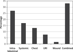
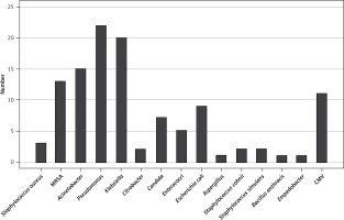
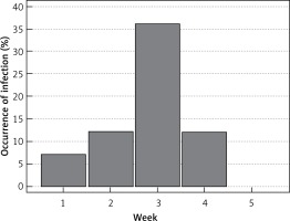
The study included 100 HCV patients who underwent LDLT (90 male and 10 female patients). Their age ranged between 20 to 63 years, with a mean ± SD of 48.42 ±7.38 years. In the entire study population, the incidence of early infectious complications was 67%. Occurrence of infection was most common in postoperative week 3 (Figure 1). The infection site was most commonly intra-abdominal (Figure 2). Figure 3 shows isolated organisms from patients with infection. Almost 50% of infections were bacterial.
Figure 1
Histogram showing the frequency of early infectious complications according to time of occurrence
Viral infections represent patients with CMV disease conforming to diagnostic criteria above. Preoperative serology for CMV was positive for IgG in 97% of cases, and positive for IgM in 10% of cases. The latter group received oral valganciclovir prophylaxis for the first 2 postoperative weeks. Donor serology for CMV was mostly (90%) IgG positive. The 3 recipients with preoperative negative serology received CMV negative donors. CMV infection without disease was not recorded as protocol surveillance cultures now in action were yet to be applied during the duration of the study. Most patients with CMV had associated bacterial or fungal infections (10 out of 11). Fungal infections were mainly Candida (9 cases) and one patient had Aspergillosis (Table I).
Association with mortality
Early infectious complications were associated with a significantly higher mortality rate in the study cohort, 34 (50.7%) patients in the infection group versus 11 (33.3%) patients in those without infections.
Logistic regression
Total leucocyte count, total bilirubin, early biliary complications, fever and CRP were found to be significant predictors of early infectious complications after LDLT. Early biliary complications included leak, stricture or both. B coefficients of significant variables are shown in Table II.
Table II
Multivariable logistic regression model showing significant predictors of early post-operative infections
| Variable | Coefficient | Std. error | P-value |
|---|---|---|---|
| TLC | –0.58 | 0.25 | 0.020 |
| Total bilirubin | 0.21 | 0.10 | 0.032 |
| Biliary complications | 5.01 | 2.53 | 0.047 |
| Fever | 9.97 | 3.84 | 0.0095 |
| CRP | 0.33 | 0.14 | 0.014 |
The incidence of early biliary complications was 22 (32.8%) patients in the group with infections (12 leaks, 6 strictures and 4 combined leak and stricture). The other group had a total of 4 (12%) patients with early biliary complications (3 leaks, 1 stricture).
The mere presence of fever of any degree was found significant, rather than the temperature value. Accordingly, it was considered as a risk factor regardless of the absolute temperature value.
ROC curve analysis of predictors
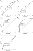
All 5 variables showed significant P value for AUC except for TLC (Table III). They showed fair sensitivity and specificity in prediction of infection. The highest AUC was that of CRP; with the criterion 24 mg/l it had 67% sensitivity and 97% specificity. The optimum cut-off value for total bilirubin was 4.4 mg/dl, with 60% sensitivity and 67% specificity. Similarly, the cut-off value for TLC was 11 × 109/l, with 42% sensitivity and 88% specificity. The presence of fever predicted infection with a high sensitivity and specificity (88% for both), while presence of early biliary complications had 34% sensitivity and 88% specificity (Figure 4).
Table III
Results of ROC curve analyses of predictor variables and risk index
| Variable | AUC | SEa | 95% CIb | Significance level pc |
|---|---|---|---|---|
| TLC | 0.530 | 0.0580 | 0.427–0.630 | 0.6096 |
| Total bilirubin | 0.655 | 0.0583 | 0.553–0.747 | 0.0079 |
| Biliary complications | 0.611 | 0.0411 | 0.508–0.707 | 0.0068 |
| Fever | 0.880 | 0.0351 | 0.799–0.936 | < 0.0001 |
| CRP | 0.901 | 0.0294 | 0.825–0.952 | < 0.0001 |
| Risk Index | 0.905 | 0.0314 | 0.830–0.955 | < 0.0001 |
Derivation of the risk index
The authors decided to combine the above predictors into a screening tool with higher sensitivity for prediction of infection. Also, CRP had the highest sensitivity, but at a relatively high cut-off value of 24 mg/l. This value is too late after the onset of infection, with little benefit concerning prompt management of high risk patients. Using a lower CRP value will result in its poor performance if used on its own: A cut-off CRP value of 10 mg/l has around 96% sensitivity, while specificity falls to 56%. Multiple combinations were tested in order to derive the most efficient index while keeping it user friendly. Cut-off values should be as low as possible to aid in early diagnosis. The CRP was successfully combined with other predictors at a cut-off value of 10 mg/dl with improved sensitivity and specificity. Optimum cut-off values for TLC and total bilirubin as derived from ROC curves were satisfactory.
For each patient, the risk index was calculated by adding a score of 1 point for every significant predictor. One point was scored for the presence of fever and for the presence of biliary complications. One point was scored for total bilirubin ≥ 4 mg/dl, TLC ≥ 11 × 109/l and CRP ≥ 10 mg/l, respectively. As mentioned above, weighted scores according to respective odds ratio were not used as they provided no additional statistical advantage to the index; therefore, the priority was to keep it simplified.
Comparison of ROC curves
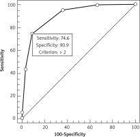
Figure 5 shows the ROC curve analyses for the risk index. The risk index predicted infection with the highest sensitivity and specificity as compared with each predictor on its own. AUC was also superior, 0.91 with 95% CI 0.830–0.955 and significance level < 0.0001, as shown in Table III. Table IV shows comparison of the risk index ROC curve against each predictor separately.
Table IV
Comparison of ROC curve analyses of each predictor variable versus the risk index
| Variable | Difference between areas | Standard errora | 95% confidence interval | Significance level p |
|---|---|---|---|---|
| Index vs. TLC | 0.376 | 0.0518 | 0.274–0.477 | < 0.0001 |
| Index vs. total bilirubin | 0.251 | 0.0517 | 0.150–0.352 | < 0.0001 |
| Index vs. biliary complications | 0.294 | 0.0442 | 0.208–0.381 | < 0.0001 |
| Index vs. fever | 0.0258 | 0.0333 | –0.0395 – 0.0911 | 0.4390 |
| Index vs. CRP | 0.0043 | 0.0318 | –0.0580 – 0.0666 | 0.8925 |
Discussion
The study revealed that 67 of our patients (67%) developed early post-operative infections; bacterial infection was the predominant type of infection followed by viral then fungal infections. Bacterial infection was the commonest form of infection as observed by previous studies [1, 12, 13].
The most common sites of infection described in the literature were surgical site, intra-abdominal, chest and urinary tract [14, 15]. Most of our patients had combined infection of above-mentioned sites.
In this study, the most common isolated organisms were Klebsiella, Pseudomonas, Acinetobacter and MRSA [16].
Organisms commonly isolated in other studies are Staphylococcus aureus [17, 18], Streptococcus viridians, Streptococcus pneumoniae and members the Enterobacteriaceae [19]. A study performed on 233 LTx recipients, the three most common organisms isolated in bacteremic patients were MRSA, Klebsiella pneumoniae, and Pseudomonas aeruginosa [20]. In a similar study performed on 221 LTx recipients, drug-resistant Gram-positive Staphylococcus aureus and Enterococcus (MRSA and VRE) were the main pathogens causing infection following liver transplantation [21].
The CMV was the only viral infection in our patients, although it is most common elsewhere [22].
Also, 54% of the patients with CMV acquired bacterial infections, probably enhanced by the immunomodulatory effect of the virus [23]. Lastly, 5 of 8 (62%) patients with fungal infection had Candidiasis while the 3 others (37%) had Aspergillosis, in line with similar studies [3, 24].
As observed in this study, previous literature describes a worse outcome for patients who developed post-operative infections than those who did not [1, 3, 12, 15, 25, 26]. It has been stated that around one third of mortality after liver transplantation was due to bacterial and fungal infections [27]. Mortality due to post-operative infections and sepsis was the most common cause of death even after liver resection for malignancy, in a previous study [28].
Taking into account all variables included in our regression model, TLC, total bilirubin, early biliary complications, fever and CRP were independently associated with early post-LTx infection, indicating a predictive value. With the exception of CRP, other variables were more or less fair as screening tools for infection as evaluated by ROC curve analysis. CRP alone is definitely guiding our screening for infections, but it seems to give an alarm that is “too late” according to ROC curve values. It should be highlighted that the statistical comparison of the risk index to CRP is biased. The optimum CRP cut-off value in question is 24 mg/dl, which would be a late sign for already established infection. At 10 mg/dl, CRP on its own has a low specificity for infection. Thus, CRP as low as 10 mg/dl was combined with other predictors to yield a risk index with a higher overall performance.
Similarly, TLC ≥ 11 × 109/l did not classically seem alarming on regular follow-up. The novel cut-off point suggested by this study should change our interpretation of this routine laboratory marker. The role of such a predictor to be an effective component of the risk index again serves as an “early alarm” for high risk patients.
A total bilirubin ≥ 4 and the presence of early biliary complications seem to be two related factors. However, bilirubin elevation is associated with an increased risk of infection independent from biliary complications, as evidenced by multivariable regression analysis. Each of the two factors can impose an elevation in the risk index for infection.
Comparing ROC curves proved the index to be superior to any of the predictors on its own. On a closer look at the coordinates of the ROC curve (Table V), the authors consider an index value of 1 as “at risk” of infection. Index values of 2 or 3 indicate that the patient is “at high risk” of infection. Values of more than 3 indicate “very high risk” or established infection, as 100% of such patients developed post-operative infection. A risk index summary is shown in Table VI.
Table V
Criterion values and coordinates of the ROC curve for the risk index
Table VI
Summary of clinical significance of the risk index
| Index | Risk of infection |
|---|---|
| 0 | No risk |
| 1 | At risk |
| 2–3 | At high risk |
| > 3 | Very high risk |
In the setting of liver transplantation, the interference of immunosuppressive therapy with the classical humoral manifestations of infection renders the early diagnostic capacity of infections a major challenge. Novel markers are present and under way; however, the need of infection surveillance throughout the early post-operative period calls for relatively cheap markers that are readily available, even several times a day. The proposal of a combined score of easily obtained humoral parameters totally fits this requirement. All necessary laboratory parameters are already taken as a routine, at least once daily, in standard liver transplantation protocols, with no additional costs required.
The risk index can be used for detection of patient subsets at higher risk of infection with no additional cost in such routinely taken parameters. Although components of the risk index are classically associated with infections, this study suggests new lower cut-off points that should be encountered earlier in the course of infections, probably before they are clinically evident. The authors recommend that it should be done on a daily basis starting from day 1, in order to catch its initial rise and define the risk category. Risk-stratified preventive, diagnostic and therapeutic strategies for early post-LTx infection should be developed within the center protocols.
Needless to point out, this was the first cohort of patients which was used to derive the index as described. The risk index needs to be further validated by the next cohort of patients in our center as well as in other centers. Also, the efficacy of additional measures in improving the outcome of high risk patients need to be measured in future studies.
In conclusion, early post-operative infections are the leading cause of morbidity and mortality after liver transplantation. The presence of at least 3 of the following can predict infections with 76% sensitivity and 91% specificity: TLC ≥ 11 × 109/l, total bilirubin ≥ 4, early biliary complications, fever and CRP ≥ 10. The use of a combined risk index may help in earlier identification of high risk patients; thus prompt risk-stratified management plans may be initiated to improve the outcome of LDLT.


