Introduction
Estimation of fetal weight (EFW) is a standard ultrasonographic procedure in antenatal care. It is one of the crucial parameters for adequate planning and managing the time and route of delivery. It helps to detect fetal growth abnormalities and determine whether elective caesarean section is indicated if fetal macrosomia is suspected. The investigators developed several regression equations to perform this estimation. At least 30 formulas for fetal weight estimation have been published [1]. These formulas are not as precise as actual birth weight (BW) and they are typically associated with estimation errors. Many of them are incorporated in commercially available ultrasound software. Formulas for calculating EFW most commonly are a combination of two-dimensional measurements (i.e. biparietal diameter (BP), head circumference (HC), abdomen circumference (AC), and femur length (FL)) and many factors potentially affect the accuracy of the estimation (i.e. race, maternal adiposity, amniotic fluid index, fetal abnormalities, sex and gestational age) [2]. The prediction of fetal weight by formulas based on three-dimensional ultrasonography (3DUS) measurements is relatively new. Fetal soft tissue parameters – fractional limb volumes: arm (AVol) and thigh (TVol) – are most commonly employed. Multiple available regression equations with other two- and three-dimensional parameters may be combined [3–5]. For the purpose of this study, we employed the least complicated – a single parameter Lee formula. It calculates EFW with TVol. The regression equation is as follows: EFW = e (4.708 + 0.7596 × ln(TVol)), where e is Euler’s number (e = 2.71828) [6]. The objective of this study was to evaluate the Lee formula, which is based on TVol, in the daily practice of estimating fetal weight before delivery. We compared it to one of most commonly used formulas, the Hadlock I formula [7] (log10 EFW = 1.3596 – 0.00386 AC × FL + 0.0064 × HC + 0.00061 BPD × AC + 0.0424 × AC + 0.174 × FL), combined with four measurements: BP, HC, AC, and FL.
Material and methods
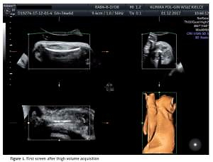
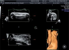
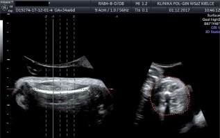
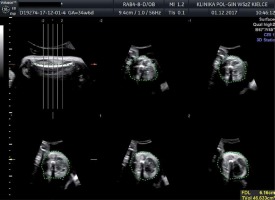
In this study we included patients in singleton, term pregnancy with cephalic presentation of the fetus. All of the patients volunteered for delivery in our clinic and consented to undergo ultrasound examination and participate in this study. Exclusion criteria were pre-labour rupture of membranes and lack of consent for participation in the study. One hundred and four singleton pregnant women met the inclusion criteria. Gestational age was between 37 and 41 (median: 39 weeks) based on the first day of the last normal menstrual period. Patients prospectively underwent three-dimensional ultrasonography for estimating TVol and two-dimensional fetal measurements with BP, HC, AC and FL taken during the same examination. Thigh volume measurement was obtained by a sagittal sweep that included both ends of the femoral diaphysis during maternal breath-holding (Figure 1). Partial volume (50% of femoral length) was automatically subdivided into five equidistant slices that were centred along the mid-thigh (Figure 2), then slices were traced manually from the transverse view of the extremity to obtain TVol (Figure 3). The Lee formula was used to calculate EFW. Evaluation took place within 3 days of delivery and was done by one certificated ultrasonographer. An independent observer measured the time taken to perform the necessary measures in 20 randomly selected patients. Amniotic fluid index (AFI) was also estimated in each patient. Women were recruited in the Department of Obstetrics and Gynaecology, Provincial Combined Hospital in Kielce. We used GE Healthcare Voluson E8 with three-dimensional curved-array abdominal transducer RAB 4–8 D and software to implement the Lee and Hadlock formulas. Immediately after delivery, neonatal staff measured BW. The percentage error (PE) between EFW and BW was calculated using the equation PE = (EFW – BW/BW) × 100%, then mean percentage errors (MPE) were calculated for each formula separately. Absolute percentage error (APE = |EFW – BW|/BW) × 100%) and median absolute percentage errors (MAPE) were calculated. We compared the MPEs and MAPEs of the formulas. We also compared the proportion of newborns with estimated BWs within ± 5% and ± 10% of actual BW. We calculated statistical measures of test performance in detecting fetal macrosomia (arbitrarily set at 4000 g) (Table I).
Table I
Comparison of formulas according to statistical analysis
| Parameter | Lee formula | Hadlock formula |
|---|---|---|
| MPE (± SD) (p < 0.05) | 2.13 ±9.31% | –2.02 ±8.79% |
| MAPE (Q1–Q3) (p = 0.56) | 6.09% (2.53–10.7) | 6.10% (3.43–10.69) |
| Proper estimation within ±10% BW, (%) (p = 0.11) | 73 | 71 |
| Proper estimation within ±5% BW (p < 0.05) | 33% | 42% |
| Time taken for measurements (p = 0.16) [s] | 69 | 58 |
| Sensitivity* | 85% | 60% |
| Specificity* | 88% | 96% |
| Positive predictive value* | 62% | 80% |
| Negative predictive value* | 96% | 90% |
| Accuracy* | 88% | 89% |
Results
Mean BW in the studied population was 3504 g (±575 g) and ranged between 2220 g and 4890 g. Distribution of fetal weight tended toward a normal distribution. The percentage of newborns with weight over 4000 g was 19.2% (20 fetuses). Median AFI was 14, and there was no case of oligohydramnios defined as AFI < 5 cm. MPEs of the Lee formula and Hadlock formula were statistically significantly different (2.13 ±9.31% vs. –2.02 ±8.79%, p = 0.001, power = 0.93 for α = 0.05). MAPEs of formulas were 6.09% for Lee and 6.10% for Hadlock and were not significantly different (p = 0.56). The proportion of newborns with estimated BWs within ±10% of actual BW was not significantly different between formulas (73% vs. 71%, p = 0.11). There was a significant difference in the proportion of the newborns with estimated BWs within ±5% (33% vs. 42%, p = 0.000006). Statistical measurements for test performance in detecting fetuses with BW ≥ 4000 g were, for the Lee formula, sensitivity 85% specificity 88% accuracy 88%; in our population positive predictive value (PPV) was 62% and negative predictive value (NPV) was 96%. Hadlock formula test performance was sensitivity 60%, specificity 96%, accuracy 89%, PPV 80%, NPV 90%. Mean time of taking necessary measurements for the Hadlock I formula was 58 s compared with 69 s for taking measurements for the Lee formula. There was no significant difference between groups (p = 0.16).
Discussion
Human newborn infant’s fat mass constituted 14% of birth weight but contributed to 46% of its variance [8]. Intrauterine growth abnormalities especially influence this compartment of the fetal body. The investigators demonstrated a correlation between 3DUS fetal limb volume (AVol, TVol) and birth weight [4, 6, 9]. Thigh volume was easily and rapidly measured, and highly reproducible among blinded observers [4]. It is incorporated in many formulas for the estimation of fetal weight, which contain other fetal measurements such as AC, BPD, AVol. These were reported and prospectively validated by the investigators [4, 10]. The advantage of the equation that we chose is that it is a single parameter model with a shorter procedure time. From the reported literature, the time taken to conduct the measurement was 1 to 2 min with manual tracing of the slices [4]. This is similar to our results. For the purpose of shortening the measurement time, software for the automatic tracing of slices was investigated with almost total correlation with the original manual tracing method (0.993 to 0.998) [11]. In our study, the Lee formula tended to over-estimate BW. This result has higher sensitivity in detecting macrosomic fetuses, but lower specificity than the Hadlock formula, and, because of the issue of over-estimating BW, it had an improved negative predictive value in our population. We chose 4000 g as the cut-off because the risk of shoulder dystocia increases over that cut-off [10, 11]. In our study, the Hadlock formula was superior in the estimation within 5% BW, but there was no difference in the estimation within 10% BW. In our opinion, differences in the accuracy of estimation within 5% BW are less clinically significant before delivery than 10% BW estimation. The disadvantage of the Hadlock formula when the fetal head is advanced in the birth canal is the difficulty or even impossibility of acquiring a good plane for the measurement of fetal HC and BPD. Since cephalic presentation represents 97% of all fetuses at term [12–14], TVol seems to be a reasonable alternative to formulas in which head measurements are included. Each patient in our study had intact fetal membranes and there were no cases of oligohydramnios (defined by amniotic fluid index < 5 cm). Further evaluation needs to be done to assess the usefulness of TVol measurement before labour in cases of rupture of the membranes or oligohydramnios since the fluid layer helps to differentiate soft tissue borders from adjacent fetal limb and uterine wall and a large volume of amniotic fluid helps in 3DUS evaluation. Several technical considerations need be discussed here about TVol measurement. Excessive abdominal transducer pressure can decrease the layer of amniotic fluid, which helps in TVol measurement, movement artefacts can be minimized by maternal breath holding, and the sweep angle is particularly relevant for macrosomic fetuses and, in the case of term pregnancy, it should be approximately 85°. Compression by adjacent structures may be another consideration and this is more likely to occur with decreased amniotic fluid volume. There is also the issue of inadvertent confusion between the fetal arm and leg, and that can be minimized if transducer orientation is correctly oriented in relation to fetal lie [15, 16].
In conclusion, TVol measurement incorporated in the Lee single parameter formula was comparable to the Hadlock I formula in accuracy in predicting fetal weight before delivery. There was no significant difference in the time needed for taking necessary measurements between two groups. The Lee formula could be useful in cases when the fetal head is engaged in the maternal pelvis and taking AC and BPD measurements is impossible. Further evaluation is needed to assess the usefulness of this formula in specific obstetrical situations.


