Current issue
Archive
Manuscripts accepted
About the Journal
Editorial office
Editorial board
Section Editors NEW
Abstracting and indexing
Subscription
Contact
Ethical standards and procedures
Most read articles
Instructions for authors
Article Processing Charge (APC)
Regulations of paying article processing charge (APC)
INTERNAL MEDICINE AND GERIATRICS / STATE OF THE ART PAPER
Analysis of the current status of computed tomography diagnosis of sarcopenia
1
Department of Endocrinology, Union Hospital, Tongji Medical College, Huazhong University of Science and Technology, Wuhan, China
Submission date: 2023-11-15
Final revision date: 2024-07-15
Acceptance date: 2024-07-16
Online publication date: 2024-07-25
Corresponding author
Wenfang Xia
Department of Endocrinology Union Hospital Tongji Medical College Huazhong University of Science and Technology 430022 Wuhan, China
Department of Endocrinology Union Hospital Tongji Medical College Huazhong University of Science and Technology 430022 Wuhan, China
KEYWORDS
TOPICS
ABSTRACT
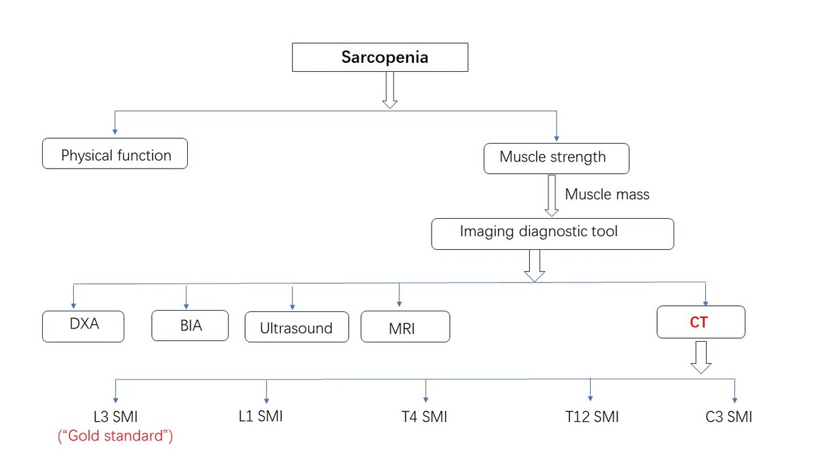
Sarcopenia is a clinical syndrome characterized by the reduction of skeletal muscle mass and strength, leading to adverse events such as falls, fractures, frailty, disability, and increased mortality. Compared to previous diagnostic techniques such as dual-energy X-ray absorptiometry (DXA), bioelectrical impedance analysis (BIA), and body composition analysis, computed tomography (CT) offers significant advantages. Opportunistic CT imaging, enhanced by artificial intelligence (AI) software, provides a superior diagnostic tool for sarcopenia. AI software can automatically segment muscle groups on opportunistic CT images from different populations, enabling the efficient calculation of body composition parameters and more accurate and rapid diagnosis of sarcopenia. Early intervention may significantly reduce adverse clinical outcomes associated with sarcopenia. This study aims to evaluate the advantages of using CT images compared to traditional diagnostic techniques and to assess the value of skeletal muscle parameters at different spinal levels on opportunistic CT images for diagnosing sarcopenia.
REFERENCES (90)
1.
Truswell AS. Standardization of nomenclature of body composition in weight loss. Am J Clin Nutr 1998; 67: 492-3.
2.
Cao M, Lian J, Lin X, et al. Prevalence of sarcopenia under different diagnostic criteria and the changes in muscle mass, muscle strength, and physical function with age in Chinese old adults. BMC Geriatr 2022; 22: 889.
3.
Chew STH, Tey SL, Yalawar M, et al. Prevalence and associated factors of sarcopenia in community-dwelling older adults at risk of malnutrition. BMC Geriatr 2022; 22: 997.
4.
Keevil VL, Romero-Ortuno R. Ageing well: a review of sarcopenia and frailty. Proc Nutr Soc 2015; 74: 337-47.
5.
Uchmanowicz I, Rosano G, Piepoli M, et al. The concurrent impact of mild cognitive impairment and frailty syndrome in heart failure. Arch Med Sci 2023; 19: 912-20.
6.
Cruz-Jentoft AJ, Baeyens JP, Bauer JM, et al.; European Working Group on Sarcopenia in Older P. Sarcopenia: European consensus on definition and diagnosis: report of the European Working Group on Sarcopenia in Older People. Age Ageing Jul 2010; 39: 412-23.
7.
Fielding RA, Vellas B, Evans WJ, et al. Sarcopenia: an undiagnosed condition in older adults. current consensus definition: prevalence, etiology, and consequences. International Working Group on Sarcopenia. J Am Med Directors Assoc 2011; 12: 249-256.
8.
Vitale JA, Messina C, Albano D, et al. Appendicular muscle mass, thigh intermuscular fat infiltration, and risk of fall in postmenopausal osteoporotic elder women. Gerontology 2021; 67: 415-24.
9.
Cruz-Jentoft AJ, Landi F, Schneider SM, et al. Prevalence of and interventions for sarcopenia in ageing adults: a systematic review. Report of the International Sarcopenia Initiative (EWGSOP and IWGS). Review. Age Ageing 2014; 43: 748-59.
10.
Reiss J, Iglseder B, Alzner R, et al. Consequences of applying the new EWGSOP2 guideline instead of the former EWGSOP guideline for sarcopenia case finding in older patients. Age Ageing 2019; 48: 719-24.
11.
Bianchi L, Maietti E, Abete P, et al.; Investigators GG. Comparing EWGSOP2 and FNIH sarcopenia definitions: agreement and 3-year survival prognostic value in older hospitalized adults: the GLISTEN Study. J Gerontol A Biol Sci Med Sci 2020; 75: 1331-7.
12.
Jing X, Tan L, Fu H, Yang L, Yang M. Associations of ADL disability with trunk muscle mass and muscle quality indicators measured by opportunistic chest computed tomography imaging among older inpatients. Front Med 2021; 8: 743698.
13.
Lopez PD, Nepal P, Akinlonu A, et al. Low skeletal muscle mass independently predicts mortality in patients with chronic heart failure after an acute hospitalization. Cardiology 2019; 142: 28-36.
14.
Touban BM, Pavlesen S, Smoak JB, et al. Decreased lean psoas cross-sectional area is associated with increased 1-year all-cause mortality in male elderly orthopaedic trauma patients. J Orthop Trauma 2019; 33: e1-7.
15.
Tan L, Ji G, Bao T, Fu H, Yang L, Yang M. Diagnosing sarcopenia and myosteatosis based on chest computed tomography images in healthy Chinese adults. Insights Imaging 2021; 12: 163.
16.
Beetz NL, Geisel D, Maier C, et al. Influence of baseline CT body composition parameters on survival in patients with pancreatic adenocarcinoma. J Clin Med 2022; 11: 2356.
17.
Cruz-Jentoft AJ, Bahat G, Bauer J, et al.; Extended Grp E. Sarcopenia: revised European consensus on definition and diagnosis (vol 48, pg 16, 2019). Correction. Age Ageing 2019; 48: 601.
18.
Chen Z, Li WY, Ho M, Chau PH. The prevalence of sarcopenia in Chinese older adults: meta-analysis and meta-regression. Nutrients 2021; 13: 1441.
19.
Chen LK, Liu LK, Woo J, et al. Sarcopenia in Asia: Consensus Report of the Asian Working Group for Sarcopenia. Review. J Am Med Directors Assoc 2014; 15: 95-101.
20.
Wang W, Richards G, Rea S. Hybrid data mining ensemble for predicting osteoporosis risk. Conf Proc IEEE Eng Med Biol Soc 2005; 2006: 886-9.
21.
Jimenez-Gutierrez GE, Martinez-Gomez LE, Martinez-Armenta C, Pineda C, Martinez-Nava GA, Lopez-Reyes A. Molecular mechanisms of inflammation in sarcopenia: diagnosis and therapeutic update. Cells 2022; 11: 2359.
22.
Herda AA, Cleary CJ. Agreement between multifrequency BIA and DXA for assessing segmental appendicular skeletal muscle mass in older adults. Aging Clin Exp Res 2022; 34: 2789-95.
23.
Cheng KY, Chow SK, Hung VW, et al. Diagnosis of sarcopenia by evaluating skeletal muscle mass by adjusted bioimpedance analysis validated with dual-energy X-ray absorptiometry. J Cachexia Sarcopenia Muscle 2021; 12: 2163-73.
24.
Vasilevska Nikodinovska V, Ivanoski S. Sarcopenia, more than just muscle atrophy: imaging methods for the assessment of muscle quantity and quality. Rofo 2023; 195: 777-89.
25.
Radwan RW, Tang AM, Beasley WD. Computed tomography as a first-line investigation for elderly patients admitted to a surgical assessment unit. Ann R Coll Surg 2018; 100: 285-9.
26.
Lenchik L, Lenoir KM, Tan J, et al. Opportunistic measurement of skeletal muscle size and muscle attenuation on computed tomography predicts 1-year mortality in medicare patients. J Gerontol A Biol Sci Med Sci 2019; 74: 1063-9.
27.
Boutin RD, Lenchik L. Value-added opportunistic CT: insights into osteoporosis and sarcopenia. AJR Am J Roentgenol 2020; 215: 582-94
28.
Chianca V, Albano D, Messina C, et al. Sarcopenia: imaging assessment and clinical application. Abdom Radiol (NY) 2022; 47: 3205-16.
29.
Albano D, Messina C, Vitale J, Sconfienza LM. Imaging of sarcopenia: old evidence and new insights. Eur Radiol 2020; 30: 2199-208.
30.
Boutin RD, Houston DK, Chaudhari AS, Willis MH, Fausett CL, Lenchik L. Imaging of sarcopenia. Radiol Clin North Am 2022; 60: 575-82.
31.
Kaplan SJ, Pham TN, Arbabi S, et al. Association of radiologic indicators of frailty with 1-year mortality in older trauma patients: opportunistic screening for sarcopenia and osteopenia. JAMA Surg 2017; 152: e164604.
32.
Romanowski KS, Fuanga P, Siddiqui S, Lenchik L, Palmieri TL, Boutin RD. Computed tomography measurements of sarcopenia predict length of stay in older burn patients. J Burn Care Res 2021; 42: 3-8.
33.
van Heusden HC, Swartz JE, Chargi N, et al. Feasibility of assessment of skeletal muscle mass on a single cross-sectional image at the level of the fourth thoracic vertebra. Eur J Radiol 2021; 142: 109879.
34.
Bokshan SL, Han AL, DePasse JM, et al. Effect of sarcopenia on postoperative morbidity and mortality after thoracolumbar spine surgery. Orthopedics 2016; 39: E1159-64.
35.
Zopfs D, Bousabarah K, Lennartz S, et al. Evaluating body composition by combining quantitative spectral detector computed tomography and deep learning-based image segmentation. Eur J Radiolo 2020; 130: 8.
36.
Waduud MA, Adusumilli P, Drozd M, et al. Volumetric versus single slice measurements of core abdominal muscle for sarcopenia. Br J Radiol 2019; 92: 20180434.
37.
Amini B, Boyle SP, Boutin RD, Lenchik L. Approaches to assessment of muscle mass and myosteatosis on computed tomography: a systematic review. J Gerontol A Biol Sci Med Sci 2019; 74: 1671-8.
38.
Jovanovic N, Chinnery T, Mattonen SA, Palma DA, Doyle PC, Theurer JA. Sarcopenia in head and neck cancer: a scoping review. PLoS One 2022; 17: e0278135.
39.
Lin TY, Chen YF, Wu WT, et al. Impact of sarcopenia on the prognosis and treatment of lung cancer: an umbrella review. Discov Oncol 2022; 13: 115.
40.
Mauro E, Diaz JM, Garcia-Olveira L, et al. Sarcopenia HIBA score predicts sarcopenia and mortality in patients on the liver transplant waiting list. Hepatol Commun 2022; 6: 1699-710.
41.
Kim DW, Ahn H, Kim KW, et al. Prognostic value of sarcopenia and myosteatosis in patients with resectable pancreatic ductal adenocarcinoma. Korean J Radiol 2022; 23: 1055-66.
42.
Derstine BA, Holcombe SA, Ross BE, Wang NC, Su GL, Wang SC. Skeletal muscle cutoff values for sarcopenia diagnosis using T10 to L5 measurements in a healthy US population. Sci Rep 2018; 8: 11369.
43.
Arayne AA, Gartrell R, Qiao J, Baird PN, Yeung JM. Comparison of CT derived body composition at the thoracic T4 and T12 with lumbar L3 vertebral levels and their utility in patients with rectal cancer. BMC Cancer 2023; 23: 56.
44.
Kim EY, Kim YS, Park I, et al. Evaluation of sarcopenia in small-cell lung cancer patients by routine chest CT. Supportive Care Cancer 2016; 24: 4721-6.
45.
Sato S, Sato M, Shinohara H. Significance of preoperative evaluation of skeletal muscle index and immune-nutritional status for patients with early-stage non-small cell lung cancer. Gen Thorac Cardiovasc Surg 2023; 71: 354-62.
46.
Liu S, Han X, Li J, et al. Feasibility of using chest computed tomography (CT) imaging at the first lumbar vertebra (L1) level to assess skeletal muscle mass: a retrospective study. PeerJ 2023; 11: e16652.
47.
Mathur S, Rodrigues N, Mendes P, Rozenberg D, Singer LG. Computed tomography–derived thoracic muscle size as an indicator of sarcopenia in people with advanced lung disease. Cardiopulm Phys Ther J 2017; 28: 99-105.
48.
Kim YS, Kim EY, Kang SM, Ahn HK, Kim HS. Single cross-sectional area of pectoralis muscle by computed tomography – correlation with bioelectrical impedance based skeletal muscle mass in healthy subjects. Clin Physiol Funct Imaging 2017; 37: 507-11.
49.
Moon SW, Choi JS, Lee SH, et al. Thoracic skeletal muscle quantification: low muscle mass is related with worse prognosis in idiopathic pulmonary fibrosis patients. Respir Res 2019; 20: 35.
50.
Kalyani RR, Corriere M, Ferrucci L. Age-related and disease-related muscle loss: the effect of diabetes, obesity, and other diseases. Lancet Diabetes Endocrinol 2014; 2: 819-29.
51.
Gao H, Wang J, Zou X, Zhang K, Zhou J, Chen M. High blood urea nitrogen to creatinine ratio is associated with increased risk of sarcopenia in patients with chronic obstructive pulmonary disease. Exp Gerontol 2022; 169: 111960.
52.
Nemec U, Heidinger B, Sokas C, Chu L, Eisenberg RL. Diagnosing sarcopenia on thoracic computed tomography: quantitative assessment of skeletal muscle mass in patients undergoing transcatheter aortic valve replacement. Acad Radiol 2017; 24: 1154-61.
53.
Wang Y, Chai L, Chen Y, et al. Quantitative CT parameters correlate with lung function in chronic obstructive pulmonary disease: a systematic review and meta-analysis. Front Surg 2022; 9: 1066031.
54.
Panthofer AM, Olson SL, Harris DG, Matsumura JS. Derivation and validation of thoracic sarcopenia assessment in patients undergoing thoracic endovascular aortic repair. J Vasc Surg 2019; 69: 1379-86.
55.
Bril SI, Wendrich AW, Swartz JE, et al. Interobserver agreement of skeletal muscle mass measurement on head and neck CT imaging at the level of the third cervical vertebra. Eur Arch Otorhinolaryngol 2019; 276: 1175-82.
56.
de Bree R, Meerkerk CDA, Halmos GB, et al. Measurement of sarcopenia in head and neck cancer patients and its association with frailty. Front Oncol 2022; 12: 884988.
57.
Chargi N, Bril SI, Smid EJ, de Jong PA, de Bree R. Cut-off values for low skeletal muscle mass at the level of the third cervical vertebra (C3) in patients with head and neck cancer. Quant Imaging Med Surg 2022; 12: 3024-33.
58.
Barnard R, Tan J, Roller B, et al. Machine learning for automatic paraspinous muscle area and attenuation measures on low-dose chest CT scans. Acad Radiol 2019; 26: 1686-94.
59.
Lenchik L, Heacock L, Weaver AA, et al. Automated segmentation of tissues using CT and MRI: a systematic review. Acad Radiol 2019; 26: 1695-706.
60.
Laino ME, Generali E, Tommasini T, et al. An individualized algorithm to predict mortality in COVID-19 pneumonia: a machine learning based study. Arch Med Sci 2022; 18: 587-95.
61.
Pella D, Toth S, Paralic J, et al. The possible role of machine learning in detection of increased cardiovascular risk patients - KSC MR Study (design). Arch Med Sci 2022; 18: 991-7.
62.
Kullberg J, Hedstrom A, Brandberg J, et al. Automated analysis of liver fat, muscle and adipose tissue distribution from CT suitable for large-scale studies. Sci Rep 2017; 7: 10425.
63.
Lee H, Troschel FM, Tajmir S, et al. Pixel-level deep segmentation: artificial intelligence quantifies muscle on computed tomography for body morphometric analysis. J Dig Imaging 2017; 30: 487-98.
64.
Weston AD, Korfiatis P, Kline TL, et al. Automated abdominal segmentation of CT scans for body composition analysis using deep learning. Radiology 2019; 290: 669-79.
65.
Polan DF, Brady SL, Kaufman RA. Tissue segmentation of computed tomography images using a Random Forest algorithm: a feasibility study. Phys Med Biol 2016; 61: 6553-69.
66.
Somasundaram E, Deaton J, Kaufman R, Brady S. Fully automated tissue classifier for contrast-enhanced CT scans of adult and pediatric patients. Phys Med Biol 2018; 63: 135009.
67.
Borrelli A, Pecoraro M, Del Giudice F, et al. Standardization of body composition status in patients with advanced urothelial tumors: the role of a CT-based AI-powered software for the assessment of sarcopenia and patient outcome correlation. Cancers 2023; 15: 2968.
68.
Herrod PJJ, Boyd-Carson H, Doleman B, et al. Quick and simple; psoas density measurement is an independent predictor of anastomotic leak and other complications after colorectal resection. Techn Coloproctol 2019; 23: 129-34.
69.
Shen DG, Wu GR, Suk HI. Deep learning in medical image analysis. Annu Rev Biomed Eng 2017; 19: 221-48.
70.
Limkin EJ, Sun R, Dercle L, et al. Promises and challenges for the implementation of computational medical imaging (radiomics) in oncology. Ann Oncol 2017; 28: 1191-206.
71.
Hsu TMH, Schawkat K, Berkowitz SJ, et al. Artificial intelligence to assess body composition on routine abdominal CT scans and predict mortality in pancreatic cancer- A recipe for your local application. Eur J Radiol 2021; 142: 109834. 4.
72.
Bedrikovetski S, Seow W, Kroon HM, Traeger L, Moore JW, Sammour T. Artificial intelligence for body composition and sarcopenia evaluation on computed tomography: a systematic review and meta-analysis. Eur J Radiol 2022; 149: 110218.
73.
Ackermans L, Volmer L, Wee L, et al. Deep learning automated segmentation for muscle and adipose tissue from abdominal computed tomography in polytrauma patients. Sensors 2021; 21: 2083.
74.
Koitka S, Kroll L, Malamutmann E, Oezcelik A, Nensa F. Fully automated body composition analysis in routine CT imaging using 3D semantic segmentation convolutional neural networks. Eur Radiol 2021; 31: 1795-804.
75.
Beetz NL, Maier C, Segger L, et al. First PACS-integrated artificial intelligence-based software tool for rapid and fully automatic analysis of body composition from CT in clinical routine. JCSM Clin Rep 2022; 7: 3-11.
76.
Just IA, Schoenrath F, Roehrich L, et al. Artificial intelligence-based analysis of body composition predicts outcome in patients receiving long-term mechanical circulatory support. J Cachexia Sarcopenia Muscle 2024; 15: 270-80.
77.
Lieffers JR, Bathe OF, Fassbender K, Winget M, Baracos VE. Sarcopenia is associated with postoperative infection and delayed recovery from colorectal cancer resection surgery. Br J Cancer 2012; 107: 931-6.
78.
McDonald ML, Diaz AA, Ross JC, et al. Quantitative computed tomography measures of pectoralis muscle area and disease severity in chronic obstructive pulmonary disease. A cross-sectional study. Ann Am Thorac Soc 2014; 11: 326-34.
79.
Liu Y, Ji F, Nguyen MH. Sarcopenia in cirrhosis: epidemiology, diagnosis, management and prognosis. Curr Opin Gastroenterol 2023; 39: 131-9.
80.
Jiang C, Wang Y, Fu W, et al. Association between sarcopenia and prognosis of hepatocellular carcinoma: a systematic review and meta-analysis. Front Nutr 2022; 9: 978110.
81.
Wang M, Tang H, Chen X, et al. Opportunistic muscle evaluation during chest ct is associated with vertebral compression fractures in old adults: a longitudinal study. J Gerontol A Biol Sci Med Sci 2024; 79: glad162.
82.
Friedman RS, Tarasova A, Jain VR, Ye K, Mansour A, Haramati LB. Predictive value of CT biomarkers in lung transplantation survival: preliminary investigation in a diverse, underserved, urban population. Lung 2023; 201: 581-90.
83.
Olmez T, Karakose E, Bozkurt H, et al. Sarcopenia is associated with increased severe postoperative complications after colon cancer surgery. Arch Med Sci 2021; 17: 361-7.
84.
Icard P, Schussler O, Loi M, et al. Pre-disease and pre-surgery BMI, weight loss and sarcopenia impact survival of resected lung cancer independently of tumor stage. Cancers 2020; 12: 266.
85.
Pekařová A, Pekař M, Daniš D, Nováková Z. CT evaluated sarcopenia signals: shorter survival for small cell lung cancer patients. Physiol Res 2021; 70 (S3): S381-6.
86.
Dong Q. Fully-automated segmentation of muscle measurement on CT in detecting central sarcopenia: a trend of standardization. Acad Radiol 2020; 27: 321-2.
87.
Rozynek M, Kucybala I, Urbanik A, Wojciechowski W. Use of artificial intelligence in the imaging of sarcopenia: a narrative review of current status and perspectives. Nutrition 2021; 89: 111227.
88.
Cespedes Feliciano EM, Popuri K, Cobzas D, et al. Evaluation of automated computed tomography segmentation to assess body composition and mortality associations in cancer patients. J Cachexia Sarcopenia Muscle 2020; 11: 1258-69.
89.
Lee YS, Hong N, Witanto JN, et al. Deep neural network for automatic volumetric segmentation of whole-body CT images for body composition assessment. Clin Nutr 2021; 40: 5038-46.
90.
Chen L, Wu F, Huang J, et al. Well-aerated lung and mean lung density quantified by CT at discharge to predict pulmonary diffusion function 5 months after COVID-19. Diagnostics 2022; 12: 2921.
Share
RELATED ARTICLE
| eISSN: | 1896-9151 |
| ISSN: | 1734-1922 |
We process personal data collected when visiting the website. The function of obtaining information about users and their behavior is carried out by voluntarily entered information in forms and saving cookies in end devices. Data, including cookies, are used to provide services, improve the user experience and to analyze the traffic in accordance with the Privacy policy. Data are also collected and processed by Google Analytics tool (more).
You can change cookies settings in your browser. Restricted use of cookies in the browser configuration may affect some functionalities of the website.
You can change cookies settings in your browser. Restricted use of cookies in the browser configuration may affect some functionalities of the website.